 |
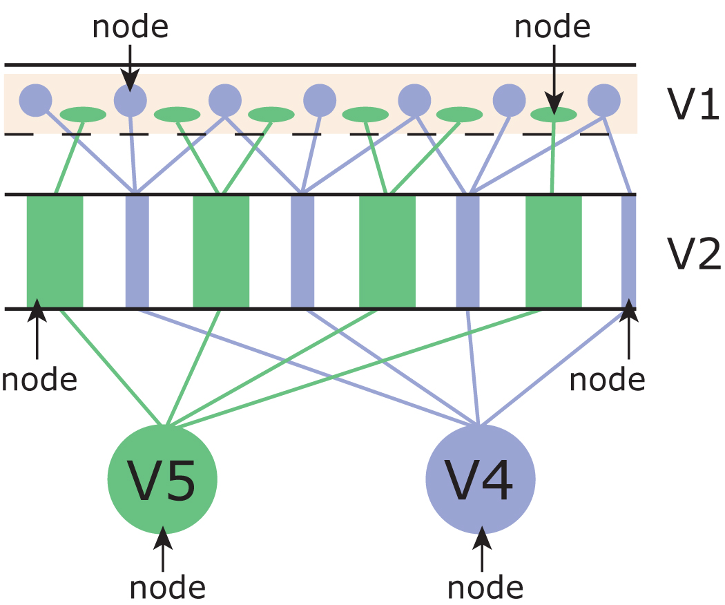
Figure 1. (Figure 13.1 in the book.) Diagrammatic illustration of nodes and essential nodes. Signals related to color (blue) and motion (green) reach distinct compartments in the primary visual cortex (V1) and the area surrounding it (V2). The specialized compartments of V1 and V2 constitute nodes that project to further nodes V4 (for color) and V5 (for motion). The latter are essential nodes in that the signals in them becomes explicit and does not necessarily need to be processed further. When V4 and V5 are destroyed the nodes in V1 and V2 become essential nodes and the subjects' perceptual capacities in color and motion now reflect the physiological capacities of cells in V1 and V2. |
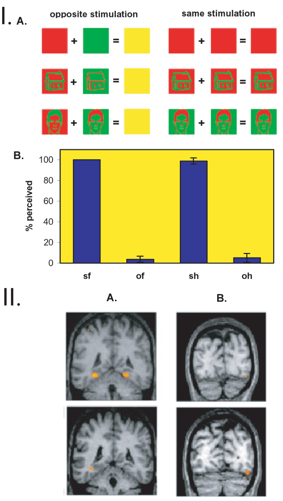 |
Figure 2 (which does not appear in the book). I. A.: A schematic
presentation of the experiment of Moutoussis and Zeki (2002). Pictures
of houses, faces, and uniformly colored controls were used. The input
to the two eyes and the expected perceptual output (Upper) and subjects'
true psychophysical performance (Lower) are shown. Continuous fusion
of the stimuli was achieved by using repetitive brief presentations.
Identical stimuli of opposite color contrast were invisible when presented
dichoptically to the two eyes (opposite stimulation), whereas identical
stimuli of the same color contrast (same stimulation) were easily
perceived. Control stimuli were never perceived either as a face or
a house. B. The averaged performance of the seven subjects in the
face/house/nothing discrimination task. The averaged percentage of
the number of stimuli perceived is shown (of a total of 448 per subject
per stimulus category) together with the standard error between the
subjects. sf, same faces; of, opposite faces; sh, same houses; oh,
opposite houses. II. Group results of brain regions showing stimulus-specific activation under conditions of same and opposite stimulation, revealing that such activation correlates with perceived and not-perceived conditions. (A Upper) The contrast same houses-same faces shows bilateral stimulus-specific activation in the parahippocampal gyrus (Talairach coordinates, 230, 244, 212 and 26, 244, 210). (Lower) The contrast opposite houses-opposite faces shows unilateral stimulus-specific activation in the same region (238, 242, 210). (B Upper) The contrast same faces-same houses reveals stimulus-specific activation in a region of the fusiform gyrus (42, 282, 212). (Lower) The contrast opposite faces-opposite houses reveals stimulus-specific activation in the same brain region (44, 274, 214). |
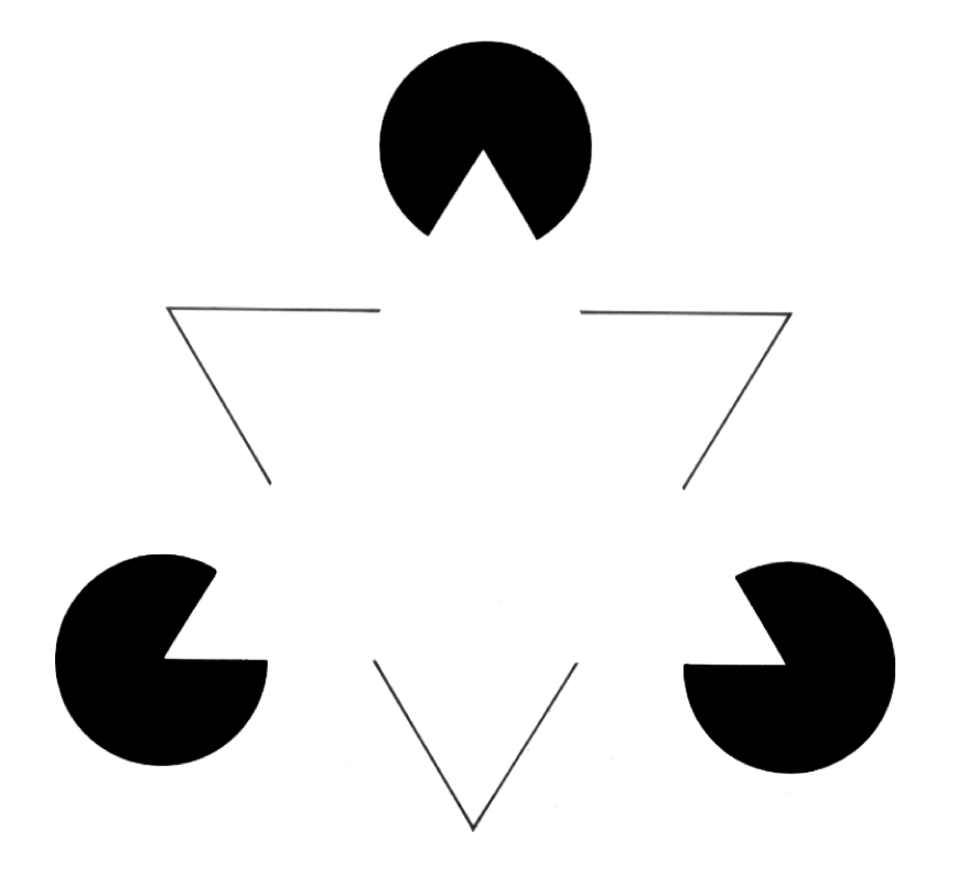
Figure 3. (Figure 13.2 in the book.) The Kanizsa triangle.
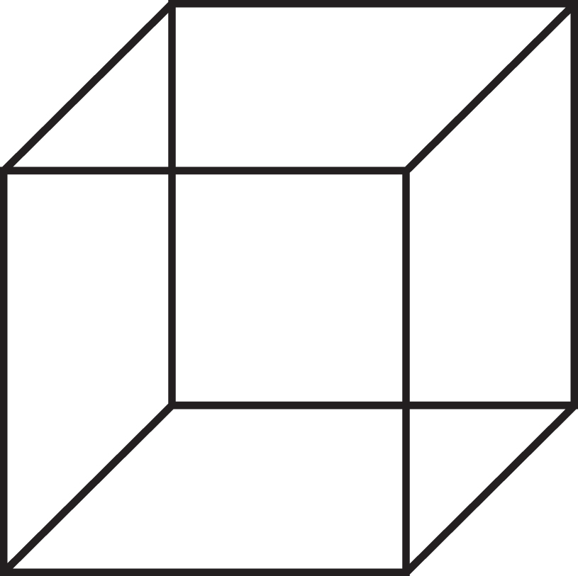
Figure 4. (Figure 13.3 in the book.) The Kanizsa cube.
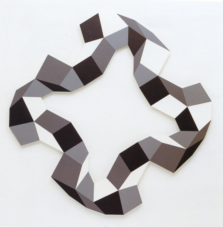
Figure 5 (which does not appear in the book). Composition
by Nathan Cohen.
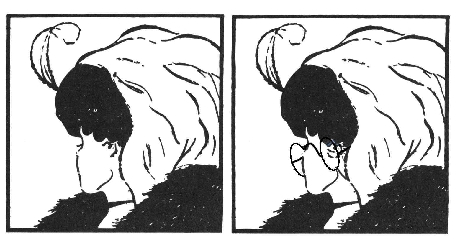
Figure 6. (Figure 13.4 in the book.) (left) Bi-stable
figure: wife/mother-in-law. (right) An attempt to dis-ambiguate the same
figure. Despite the spectacles and eyeshades to stabilize the perception
of the "mother-in-law", the figure remains unstable.
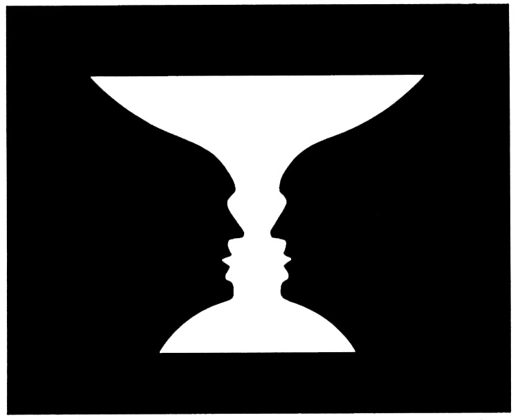
Figure 7. (Figure 13.5 in the book.) Bi-stable figure:
vase/faces.
http://www.ibiblio.org/wm/paint/auth/vermeer/i/earring.jpg
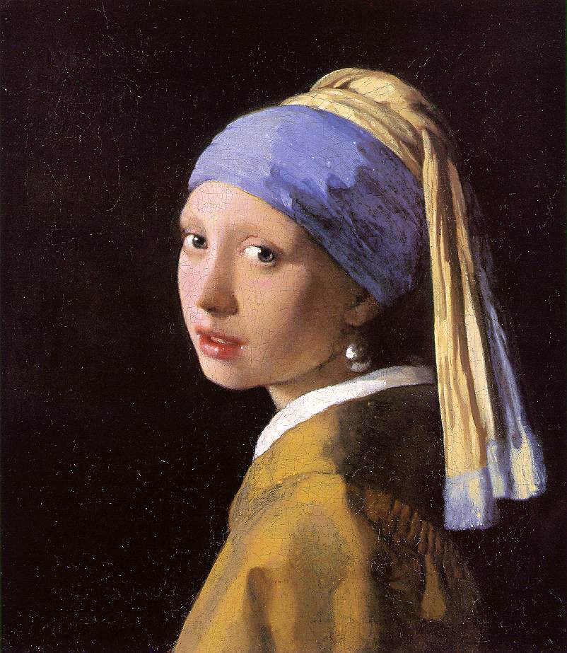
Figure 9 (which does not appear in the book). The
Pearl Earring by Johannes Vermeer (1632-75).
http://www.ibiblio.org/wm/paint/auth/vermeer/i/music-lesson.jpg
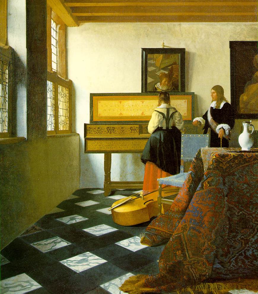
Figure 10 (which does not appear in the book). The
Music Lesson by Johannes Vermeer (1632-75).

http://www.uic.edu/depts/ahaa/classes/ah111/L18/18-19.jpg
Figure 11 (which does not appear in the book). Rondanini
Pietà by Michelangelo.
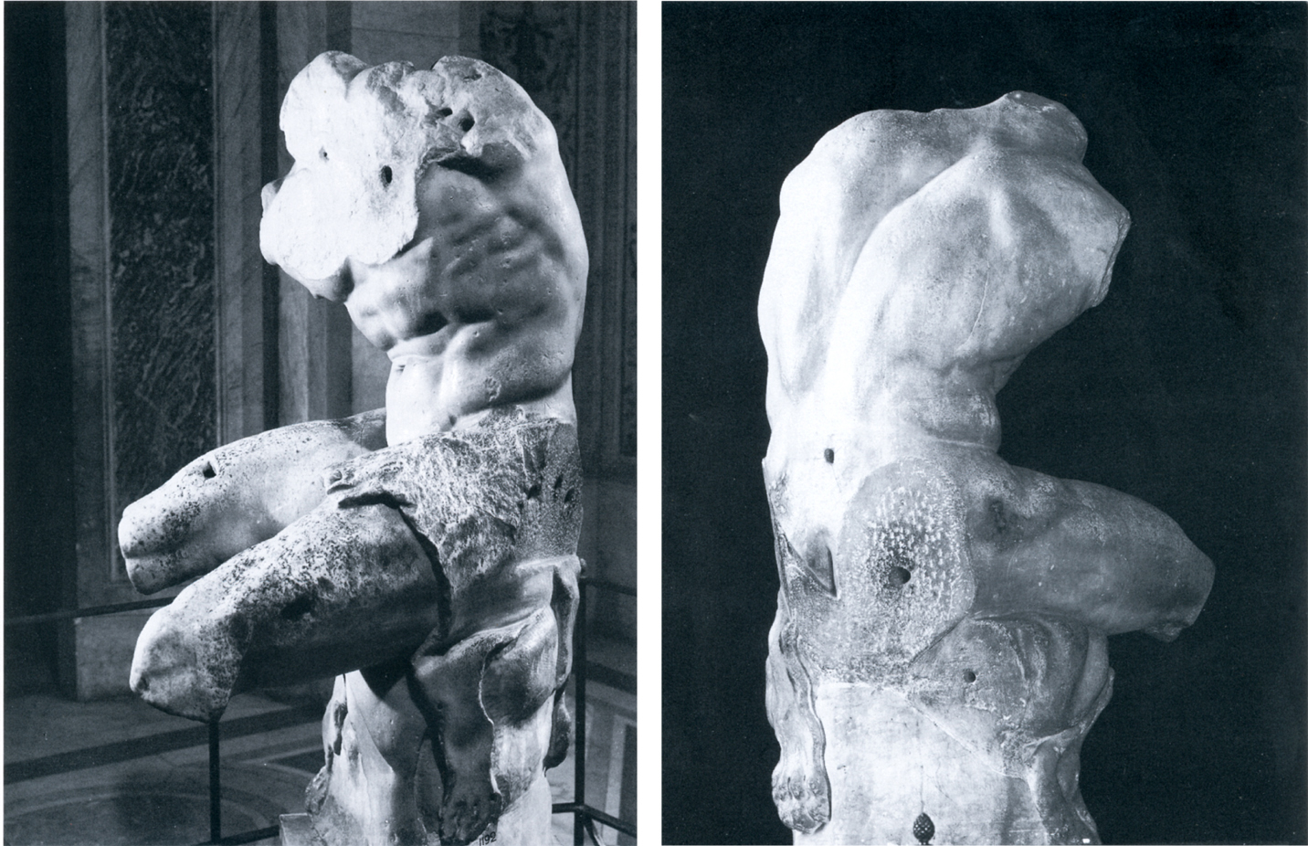
http://www.christusrex.org/www1/vaticano/SC-Torso.jpg
Figure 12 (which does not appear in the book). The
Belvedere Torso (sculptor unknown).